LISM – Laboratoire d’Ingénierie des Systèmes Macromoléculaires
The labs
- AFMB - Architecture et Fonction des Macromolécules Biologiques
- BBF - Biodiversité et Biotechnologie Fongiques
- BIAM - Bioscience and Biotechnology Institute of Aix-Marseille
- BIP - Laboratoire de Bioénergétique et Ingénierie des Protéines
- IGS - Laboratoire Information Génomique et Structurale
- IMM - Institut de Microbiologie de la Méditerranée
- iSm2 - Institut des Sciences Moléculaires de Marseille - BiosCiences
- LCB - Laboratoire de Chimie bactérienne
- LISM - Laboratoire d'Ingénierie des Systèmes Macromoléculaires
- MCT - Membranes et Cibles Thérapeutiques
- MIO - Mediterranean Institute of Oceanography - Environmental Microbiology and Biotechnology
Key Figures
- 8 Teams
- 12 Researchers
- 8 Teacher-researchers
- 13 Engineers/Technicians
- 14 Doctoral students
- 3 CDD
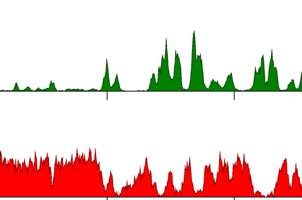
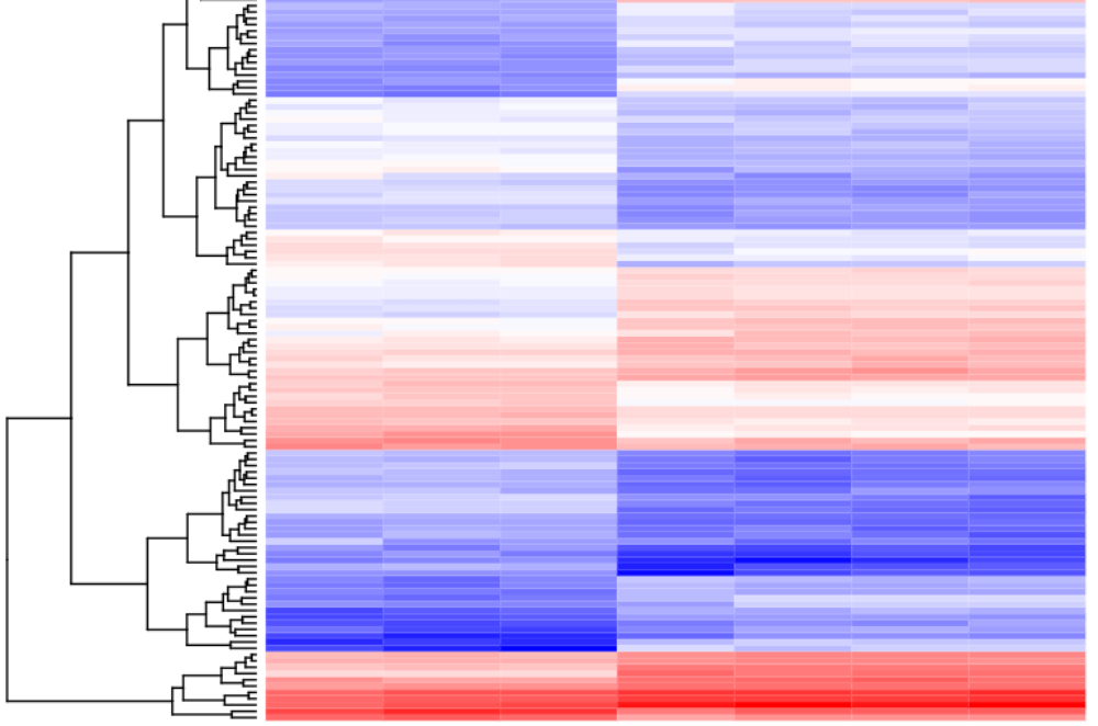
LISM brings together 8 research teams whose themes are cantered on the study of biological membranes and in particular the envelope of Gram-negative bacteria. This envelope comprises two membranes separating a periplasmic space that includes the peptidoglycan, considered to be the skeleton of the bacteria, giving it its shape and rigidité́. The envelope is the site of exchanges between the cell and its environment: entry of compounds essential to the development of the bacterium but also a protective barrier against toxic compounds. This envelope also controls the secretion of proteins in the external environment and makes it possible to detect variations in the environment and thus to regulate cellular processes to adapt to them. Finally, the inner membrane has an essential role in the cell’s energy metabolism. LISM research teams are interested in these different aspects and are internationally recognized for their work on phospholipid synthesis, signalling, transfers through the bacterial envelope (import and export) and metabolism. The techniques used are varied, from microbiology and genetics to structural biology (electron microscopy, NMR), including fluorescence microscopy and cellular microbiology.
Research
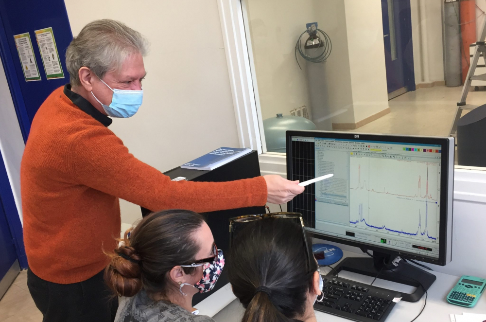
The LISM teams develop multi-scale research combining various approaches of biology, biochemistry, biophysics and structural biology in order to understand biological mechanisms from the molecular to the cellular. The LISM is particularly involved in the development of Cryo electron microscopy, a rapidly growing technique for the study of proteins and complexes. Finally, most of LISM’s themes are at the frontier of fundamental knowledge and its transfer to health applications. LISM is internationally recognized for its membrane studies, thanks to multiple collaborations in France and abroad, and to the numerous conferences given at major international congresses.
Teaching
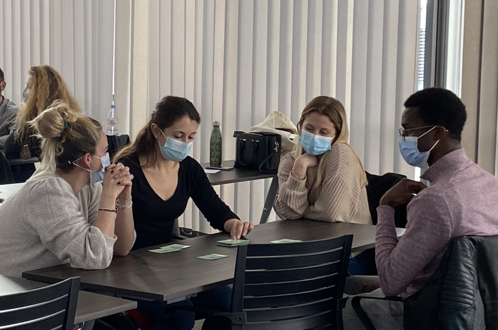
In addition to its Research activities, LISM is strongly involved in teaching and training in Biochemistry, Molecular Biology, Microbiology and Genomics. It annually provides nearly 1650 hours of teaching at AMU, as well as on other national (ENS, Collège de France, University of Toulon) and international (University of Ibadan, Nigeria) sites. In addition to the training of PhD students, LISM ensures the supervision of about twenty trainees per year (BTS, Engineering School, Bachelor and Master), and accompanies the AMU student team to the iGEM international Molecular Engineering competition (Boston, USA) since 2013. The Laboratory is also committed to social outreach and participates in various events to popularize science (science festival, presentations in schools, colleges and high schools, exhibitions and publications for the general public).
The laboratory is organised into 8 research teams each with their specific thematic.
The Teams
Assembly of bacterial multi-protein complexes – Heads: Eric Cascales
What are the players controlling the competition between bacteria within microbial communities?
The main objective of our team is to provide information on the molecular mechanisms underlying the assembly, the structure and the mode of action of multiprotein complexes associated with the cell envelope in Gram-negative bacteria, with an emphasis on secretion systems. We currently develop several lines of study on the type VI secretion system (T6SS), a contractile nanomachine that uses a spring-like mechanism to deliver effectors in target cells. We focus our efforts on understanding the regulation of the expression of T6SS gene clusters, on the structure of the apparatus, and defining how it works and how it propels effectors. An important line of study on the T6SS is to characterize effectors – their activities and targets – and how they are recruited, selected and mounted on the secretion system. A more recent project in our group wishes to define the architecture and the mechanism of action of the type IX secretion system (T9SS), a secretion system restricted to the Bacteroidetes phylum, and involved in pathogenesis and gliding motility. For all these projects, we used a combination of bacterial genetics, molecular biology, physiological tests, structural biology, and fluorescence microscopy.
Dynamics and Assembly of membrane proteins – Heads: James Sturgis
Why do membrane proteins organize and associate within membranes?
James Sturgis’ research group is interested in the dynamics and assembly of membrane proteins. We are interested in how membrane proteins fold and assemble in the membrane to form both stable structures, such as the photosynthetic apparatus, or transient dynamic structures. To investigate the assembly of membrane proteins we use a combination of different theroetical and experimental techniques. In a series of numerical approaches, we are using computer simulations and theoretical analysis to investigate the assembly process at various levels of detail (atomistic, coarse-grained and ultra-coarse grained). While at the experimental level we use spectroscopic, microscopic, molecular biology and synthetic biology methods to analyse the assembly process. We have a series of biological systems that we investigate using these techniques: AqpZ, the Bacterial photosynthetic apparatus and mitochondrial anion channel, VDAC.
Lipolysis & Bacterial Pathogenicity – Heads: Stéphane Canaan
Role of mycobacterial lipolytic enzymes and their inhibitors in host lipid uptake, persistence, virulence and antibiotic resistance
Our concern is to fight against pathogenic mycobacteria by studying lipid metabolism: identification / validation of proteins involved in lipid processes, discovery of new anti-mycobacterials (chemical, peptides) targeting lipid biosynthesis, and set up diagnostic tests related to lipid metabolism. We aim at deciphering the physiological role of lipolytic enzymes involved in lipid metabolism in pathogenic mycobacteria (such as Mycobacterium tuberculosis, M. abscessus, M. marinum, M. ulcerans), their regulation during the infection process and their impact in dormancy phase. As lipid metabolism is directly related to the viability and virulence of mycobacteria, we also develop specific models to study these enzymes and their inhibitors in order to regulate this metabolic pathway and therefore the spread of the disease. We have established that these enzymes are involved in bacterial growth, virulence (reactivation and propagation), dormancy, lipid storage and degradation as well as in cell wall biosynthesis. Moreover, our work leads to the discovery of two new families of lipolytic enzyme inhibitors with strong anti-tuberculous effects, which are useful probes to study mycobacterial lipid metabolism, thus opening a new way to fight the disease. The team expertises range from protein biochemistry (purification, biochemical characterization, expression of recombinant protein using E. coli, M. smegmatis, P. Pastoris or baculovirus expression systems), molecular and cell biology (construction of mutant strains, cell infection), lipid analysis to medicinal chemistry (organic synthesis, in silico docking, enzymology, susceptibility testing…) and cell imaging (fluorescence and electron microscopy).
Molecular transport across the bacterial cell enveloppe – Heads: Denis Duché & Laetitia Houot
How do the molecular motors of the bacterial envelope work?
Our team aims to understand the assembly and functioning of two macrocomplexes conserved in the envelope of Gram negative bacteria and involved in cell adaptation and survival: the Tol and Ton systems. Both Tol and Ton systems harness the energy produced from the proton motive force generated at the inner membrane to transduce it at the outer membrane. These complexes are involved in functions that are essential for bacterial survival. The Ton system mediates the active uptake across the outer membrane of metal, cyanocobalamin and carbohydrates. The Tol system has a more complex function, as it is involved in outer membrane integrity, in the correct localization of some polar factors and chemoreceptors, in outer-membrane phospholipid homeostasis, and in the constriction of the outer membrane during cell division. Both Tol and Ton systems can be parasitized to open a gate for bacteriotoxins (called colicins), which result in bacterial cell death. These systems are also the target of filamentous phages during the infection process, such as the M13 phage targeting E. coli, and the CTX phage which is specific of the human pathogen Vibrio cholerae. To obtain an integrate view of the inner membrane Tol and Ton complexes, we developpe an interdisciplinary study in collaboration with different laboratories, mixing genetic, biochemical and microscopy techniques.
NMR of molecular assemblies – Heads: Latifa Elantak
What are the structural bases for the assembly of protein complexes involved in cell adhesion?
Understanding the structural basis of protein assemblies that are functionally important for various aspects of cell contacts with its microenvironment. Cell contacts with the microenvironment orchestrate the development of organisms, homeostasis, and the functions of individual cells. When cells do not interact correctly or decode external molecular messages incorrectly, cellular responses are disturbed, and diseases can develop. Therefore, understanding the structural basis of protein assemblies involved in the sophisticated mechanisms of action developed by cells to interact/communicate with their microenvironment is of main importance. Our research is focused on the study of these membrane-associated protein complexes. The primary focus of our research is on how human Galectins interactions are regulated at the cell surface during cell-cell interactions. To pursue our goals, we use Nuclear Magnetic resonance (NMR) spectroscopy combined with biochemical and biophysical techniques in an integrated structural biology approach. NMR can provide an atomic-level view of the structure and dynamics of macromolecular systems in solution. A particular strength of NMR is its ability to reveal the structural details of specific interactions between biological macromolecules, even in the case of transient complexes between proteins and their targets in solution. Overall, our aim is to decipher at atomic level the assembly of multi-protein complexes, determine their structures, gain insight into their activities and regulation, and identify roles for proteins or domains of unknown function.
Pseudomonas aeruginosa pathogenicity – Heads: Sophie Bleves
Understanding the pathogenesis of Pseudomonas aeruginosa to impact its virulence
Our research team focuses towards understanding the molecular basis of virulence mechanisms of the model organism, Pseudomonas aeruginosa. P. aeruginosa is an opportunistic Gram-negative pathogen and is responsible for a wide variety of human diseases ranging from acute infections in hospital, to chronic colonization of the respiratory tract of cystic fibrosis patients. The main difficulties in treatment of patients infected with P. aeruginosa are a multi-resistance to antibiotics and a great adaptation capacity of the pathogen, which lead to the synthesis of many toxic products promoting host invasion and colonization and spread throughout the body. Antivirulence strategies that can be developed from the characterization of virulence determinants are thus key to fight against P. aeruginosa. Its virulence is multi-factorial and we particularly focus on exported or secreted products, also called effectors, which contribute greatly to P. aeruginosa virulence by facilitating colonization and survival in the eukaryotic host or competition with other bacteria. Several effectors using the Tat export pathway or the H2-T6 secretion system are under study in the lab. Characterize P. aeruginosa effectors to understand its pathogenesis and act on its virulence We investigate their characterization by multidisciplinary approaches of molecular microbiology, genetics, biochemistry and cell biology and through collaborations for microscopy and structural biology. We would like to address the mechanism of action of these effectors, their partners in the bacteria and their targets in the host.
Sensing environment & community lifestyle in Pseudomonas aeruginosa – Heads: Christophe Bordi
What are the regulatory pathways controlling biofilm formation in Pseudomonas aeruginosa?“
The “Sensing environment & community lifestyle” team is studying the regulatory pathways that control biofilm formation in P. aeruginosa as well as the processes of evolution and adaptation that this bacterium undergoes in this lifestyle. 90% of the microbiological activity in an ecosystem (aquatic environment, soil, human body, etc.) comes for the most part from sedentary flora adhering to surfaces in a structure called biofilm, which is an aggregate of micro-organisms (mostly bacteria), fixed to a surface, and stuck in a self-produced gangue of exopolymers. The presence of biofilms is increasingly identified as the recurring source of both public health and industrial problems, which are often serious and sometimes dramatic. P. aeruginosa is a gram-negative bacterium, an opportunistic human pathogen with a very high adaptive capacity since it can colonise many ecological niches. Practically harmless in healthy people, this Gram-negative environmental bacillus can become a deadly threat to immunodeficient individuals, intubated-ventilated patients in intensive care units or those suffering from chronic diseases such as cystic fibrosis. Biofilm formation monitored in flow-cell after 4 Days of growth and treats exposed to tobramycin. Dead cells were labeled red. During chromic infections, P. aeruginosa adopts a biofilm lifestyle that allows it to evade the immune system, limits the penetration of any antibiotic treatment and promotes the emergence of specific resistances. The Sensing environment & community lifestyle team studies, using genomic, fluorescent microscopy and structural biology the regulatory pathways that trigger the formation of the biofilm and the evolutionary and adaptive processes that P. aeruginosa undergoes in its lifestyle. Our aim is to propose alternative therapeutic strategies for the treatment of P. aeruginosa infections.
Virulence Nano-Macromolecular Machinosome – Heads: Eric Durand
From the intimate structure of the T6SS membrane complex to the development of virulence inhibitors targeting Gram-negative human pathogens
Our “Virulence Nano-Macromolecular Machinosome” (VN2M) team aims to Identify, to Understand, to Block and to Hijack bacterial nanomachines that challenge human health. Bacterial nanomachines play a pivotal role in bacterial life processes, for the good but also for the bad. Nanoscale dynamic protein protein interaction (PPI) drives the assembly of the myriad of protein components that build these nanomachines. Our VN2M team seeks to understand the molecular intimacy and the atomic architecture of these powerful biological nanomachines with the objective of deciphering how they work and regulate their activity. Our knowledge is then used to develop original and “chirurgical” strategies to interfere with the assembly and functioning of the virulence nanomachines, just like a “grain of sand in the nanomachine’s gears”. Our molecules or peptides will allow to combat antibiotic-resistant bacterial infections in the future. Ultimately, our increasing level of detail in the building of such “molecular cathedral” will feed our synthetic biology initiative to mimic and hijack such powerful engines for biotechnological applications, notably in cancer treatment.
Proposed Trainings


LISM contacts
LISM website- lism-dir@imm.cnrs.fr
- +33 (0)491164127
- +33 (0)491712124
-
LISM – 31 Chemin Joseph Aiguier
13402 Marseille cedex 20 – France