Microscopy Spring School – IMM
- Date: May 11-13 2026 + 1 day the next week
- Duration: 4 days
- Doctoral school equivalence: 16 hours Doctoral School equivalent
- Language: English but we will adapt the language, depending on the participants
- Name of the teachers:
- Artemis Kosta, Thierry Doan, Eric Durand , Hugo Le Guenno,
- Administrative head: Éric Pilet
- Number of students: 6 to 12
The Microscopy Spring School will include theoretical and practical courses on microscopy.
The goal of the training is for the students to have an introduction on the local microscopy facilities and techniques, as well as some lectures on more broad subjects as cutting edge structural biology and Alphafold.
Additional information
Integrative Structural Biology by Eric Durand (LCB)
Theoretical Course
Observe – Understand – Act : Structural biology (SB) provides precise observation of complex biological processes at the atomic scale. As such, SB plays a key role in the molecular understanding of pathological processes. Furthermore, by deciphering the molecular “players” involved in normal processes compared to their pathological forms, SB enables the identification of new therapeutic targets. Thus, obtaining the atomic structure of a therapeutic target is an essential step in drug development and optimization.
There are numerous SB approaches, covering a wide range of resolutions across the different scales of life. Integrative structural biology (ISB) has been developed in recent years to grasp this complexity and consolidate all this information into models. These theoretical models allow for the testing of hypotheses based on multi-parameter adjustments, which are then subjected to experimental validation.
Cryo-electron microscopy (cryo-EM) is one of the most powerful techniques for determining the structure of proteins and their complexes and to unravel the intimacy of life-important processes.
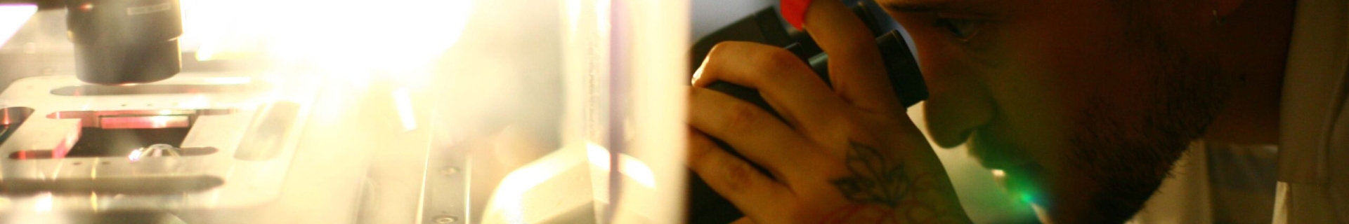
Electron Microscopy by Artemis Kosta (IMM)
We will visit the EM facilities (IMM N018), discuss the different techniques available and the questions that can be answered with these instruments. We will prepare and observe negative stained grids. We will plunge freeze grids using the Vitrobot and observe them at Cryo conditions (CryoElectron Microscopy). We will discuss strategies to obtain the answers. Key words: Transmission electron Microscopy, Negative staining, Vitrification, CryoEM
The Basics of Fluorescence Microscopy by Thierry Doan (LISM)
Using Flavobacterium johnsoniae as a thread, I will present diverse microscopy approaches that are currently used to address bacterial cell biology questions. In particular, we will go through several light microscopy modalities adapted to questions at different scales. After an introduction to the scientific questions, we will spend time to prepare samples and to discuss a few key points about fluorescence microscopy. We will then acquire fluorescence signals at different scales (colony, single cell, single molecule) and with different labels. Keywords: fluorescence microscopy, epifluorescence, oblique microscopy, protein labeling.
Confocal and 3D-SIM observation (Hugo Le Guenno)
Practical Course: Our session will begin with an introduction to the concepts of microscopy and super-resolution. Following this, we will start by capturing images using epifluorescence, then confocal microscopy and finally 3D-SIM. Through these methods, we will assess the advantages and disadvantages of each. The images generated during this session will be used later in an image analysis training session.
Image Analysis (Hugo Le Guenno)
Introduction to image processing and analysis using the free and open-source software “Fiji”. Through hands-on computer exercises in the computer lab (basement of the PH building), participants will learn to extract quantitative features from their observations. Keywords: image analysis, Fiji
Location
RdC du Bat N (Joseph Aiguier)


Facilities

Monday 11th, May 2026 10:00
Joseph Aiguier LCB